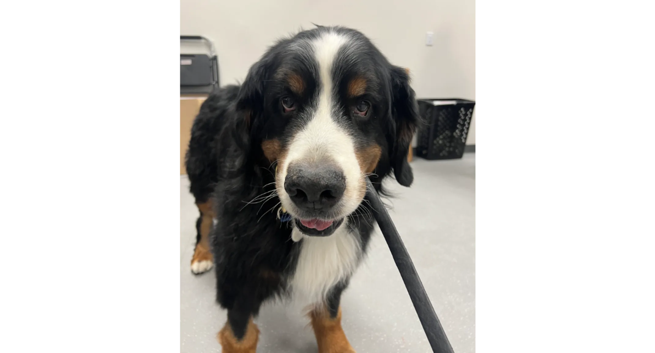
A Second Chance for Ronde: How Minimally Invasive Surgery Helped a Bernese Mountain Dog Breathe Easier
May 2, 2025 · Veterinary Services

At CASE, every patient has a story — and Ronde, an 11-year-old Bernese Mountain Dog, is no exception. When Ronde was referred to our Minimally Invasive Surgery service, he faced a serious health challenge: a suspicious lung mass in his right caudal lung lobe. But thanks to cutting-edge surgical techniques and a dedicated team, Ronde’s journey became a story of resilience, expert care, and a happy tail-wagging ending.
A Complicated Medical History
Ronde had already weathered a tough storm. Months before, he was diagnosed with primary hyperparathyroidism. During diagnostic imaging, a solitary lung mass was incidentally discovered. A CT scan revealed a 3.5 x 2.6 x 2.9 cm mass — likely a primary pulmonary tumor — in the periphery of his right caudal lung lobe. Given Ronde’s history of complications (including necrotizing fasciitis after a routine castration), his family was understandably hesitant to pursue an invasive thoracotomy.
Instead, they chose to treat his hyperparathyroidism first. A bilateral cranial parathyroidectomy and partial thyroidectomy were successfully performed by our Surgical Oncology team, and Ronde recovered smoothly with normalized calcium levels.
Watching and Waiting
While Ronde’s parathyroid condition was under control, his lung mass required ongoing monitoring. Unfortunately, follow-up thoracic radiographs showed that the mass had grown over the next four months, prompting his owners to explore other options. That’s when thoracoscopic lung lobectomy — a minimally invasive surgical alternative — became a beacon of hope.
Why Thoracoscopy?
Thoracoscopic lung lobectomy is a less invasive approach for treating smaller lung tumors (typically under 5 cm in diameter). Compared to traditional thoracotomy, this technique results in less post-operative pain, faster recovery times, and reduced trauma to the chest muscles.
After a consultation with Dr. Kyle Martin, CASE’s minimally invasive surgeon, Ronde’s owners decided to move forward with the thoracoscopic procedure.
The Surgery
A key component to the success of this surgery was the use of one-lung ventilation (OLV), made possible by an EZ-Blocker device. This technique selectively deflates one lung, giving the surgeon more space to work while minimizing trauma.
Ronde’s surgery went beautifully. The mass was removed using a thoracoscopic Endo GIA stapling device. Due to the size of the tumor, a small (4 cm) mini-thoracotomy was performed to safely extract the tissue using a specimen retrieval bag — an important step to prevent the spread of cancer cells.
The Road to Recovery
Ronde recovered without a hitch and was discharged the very next day. Just one week post-op, he was back to his usual, happy self. Histopathology confirmed the mass was a grade 1 papillary pulmonary adenocarcinoma — and it had been completely excised.
What’s Next?
Ronde’s prognosis is excellent. While the tumor was fully removed, he will continue to receive quarterly thoracic radiographs to monitor for any recurrence. With continued care and vigilance, Ronde is well on his way to many more adventures.
At CASE, we’re proud to offer advanced minimally invasive procedures that prioritize comfort, precision, and recovery. Ronde’s journey reminds us how innovation — paired with compassionate care — can truly transform lives.
